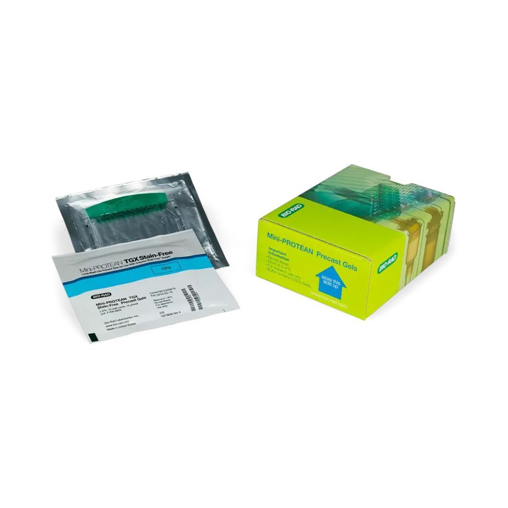
4–20% Mini-PROTEAN® TGX Stain-Free™ Protein Gels, 10 well, 50uL
Enquire for Price
- Pkg of 10, 4–20% precast polyacrylamide gel, 8.6 × 6.7 cm (W × L), 10 well, 50 µl, for use with Mini-PROTEAN Electrophoresis Cells
- Mini-PROTEAN TGX Stain-Free precast gels for polyacrylamide gel electrophoresis (PAGE) allow for fast run times, high transfer efficiency, and rapid detection of proteins with stain-free enabled imagers.
- Please contact our application specialists using our enquiry form or at sales@lasec.com for more options from our wide range of Mini-PROTEAN TGX Stain-Free Precast Protein Gels.
- Online ordering is currently limited to South African customers, but our full range is still available. If you are ordering from outside South Africa, please log an enquiry or email internationallabsales@lasec.com, and our international sales team will gladly assist you through our traditional sales channels.
Availability:On Request
SKU
BBRD4568094
BrandBio-Rad
Mini-PROTEAN TGX Stain-Free Precast Gels are based on the long-shelf-life TGX (Tris-Glycine eXtended) formulation and include unique trihalo compounds that allow rapid fluorescent detection of proteins with Bio-Rad stain-free imaging systems.
Features and Benefits of Mini-PROTEAN TGX Stain-Free Precast Gels
- Run up to 4 Mini-PROTEAN TGX Stain-Free Protein Gels using the Mini-PROTEAN Tetra Cell
- Run times as short as 15 minutes
- Transfer proteins in as little as 15 minutes; verify transfer to membrane with a stain-free enabled imager
- Inexpensive Laemmli buffer system, low running costs
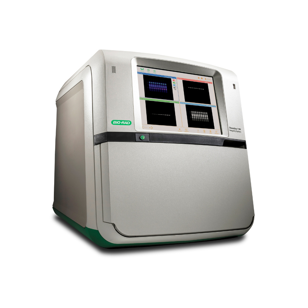
- Gel images and complete analysis in less than 5 minutes after electrophoresis
- Comparable sensitivity to Coomassie stain
- Better reproducibility and quantitation compared to staining procedures
Applications and Uses of Mini-PROTEAN TGX Stain-Free Precast Gels
- 1-D polyacrylamide gel electrophoresis for protein separation
- Accurate molecular weight estimation
- Robust system for complex samples
- One gel for western blotting, staining, and mass spectrometry
Innovative Stain-Free Technology
Mini-PROTEAN TGX Stain-Free Precast Gels do not contain SDS. For standard denaturing electrophoresis, use sample and running buffers containing SDS. To retain native protein conformation and activity, these gels can be run with a sample and running buffer that does not contain SDS.
Mini-PROTEAN TGX Stain-Free Gels retain Laemmli-like separation characteristics using standard sample and Tris/glycine running buffers. Protein migration patterns under non-denaturing conditions do not provide accurate molecular weight estimations.
The trihalo compounds in Mini-PROTEAN TGX Stain-Free Gels react with tryptophan residues in proteins in a UV-induced 1-minute reaction to produce fluorescence. The fluorophores remain covalently bound to the proteins throughout electrophoresis and blotting and may be subsequently visualized in gels or on PVDF membranes multiple times for validation in a western blotting workflow. The use of stain-free imaging also allows for the normalization of bands to the total protein on a blot, eliminating the use of housekeeping proteins or stripping and reprobing.
| Brand | Bio-Rad |
|---|
Write Your Own Review

